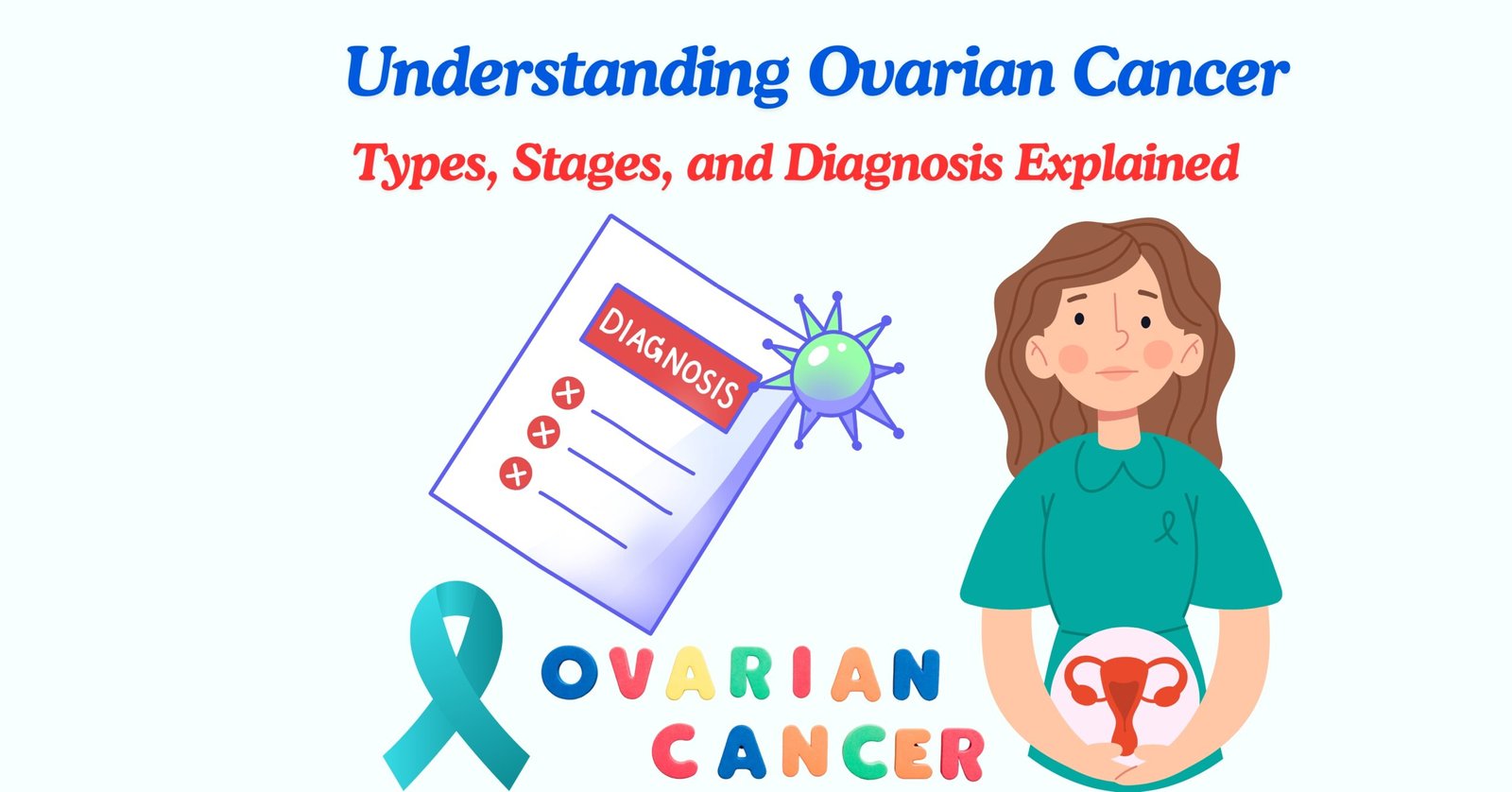
OVARIAN CANCER
Ovarian cancer is one of the most leading cause of death in women. It is an abnormal growth of cells in ovary either in the endometrium, fallopian tubes, uterus or elsewhere arises from the ovary.
Ovarian cancer is not a single disease and can be subdivided into at least five different histological subtypes that have different identifiable risk factors, cells of origin, molecular compositions, clinical features and treatments. Ovarian cancer is a global problem, is typically diagnosed at a late stage and has no effective screening strategy. Standard treatments for newly diagnosed cancer consist of cytoreductive surgery and platinum-based chemotherapy.
Sub-types of Ovarian Cancer
- Epithelial (This cancer develops in the epithelial tissue, a thin lining that covers the outside of an ovary).
- High grade serous (HGSOC) 75% (High grade grow and spread faster)
- Mucinous 10% (These tumors are more distinct and respond well to treatments. They tend to be large (around 8 inches or 20 centimeters). Typically, they only affect ovaries, (cells are large and filled with fluid), meaning that most of the tumor is made up of cells coated in mucus.).
- Endometrioid 10% (It affects the endometrium, the inner lining of uterus).
- Clear cell (Clear cell carcinoma of the ovary is a malignant epithelial tumor composed of clear, eosinophilic cells
- Low grade serous (This slow-growing cancer affects people at a younger age. Doesn’t respond well to CT).
- Germ cell (Ovarian germ cell tumor is a disease in which malignant (cancer) cells form in the germ (egg) cells of the ovary).
- Stromal Stromal cells are among the three most common cell types to be affected by ovarian cancer.
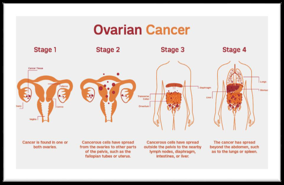
STAGES OF OVARIAN CANCER
Stage-I
- The stage I is confined only to ovary. Subtypes include IA (one ovary but no ruptured capsules or capsule is intact ) IB (Two ovaries are involved) and IC (either IA or IB with ruptured capsules).
- Stage-II
- It extends to the pelvis further categorize to subtypes including IIA -Involves fallopian tube or uterus IIB -Extension to other pelvic tissues ,IIC- Either IIA or IIB but with ruptured capsule .
- Stage-III
- It extends beyond the true pelvis IIIA Tumor limited to true pelvis but microscopic positive biopsy outside the pelvis ,IIIB Abdominal implants up to 2 cm ,IIIC -Positive lymph nodes or abdominal implants > 2 cm.
- Stage-IV
- It metastasizes to other organs of the body.
- Diagnosis:
- Blood tests: These tests will check for high levels of a marker called CA-125. (Normal 0-35 units/mL)
- Imaging tests: Examples include a transvaginal ultrasound, an MRI scan, or a CT scan.
- Laparoscopy: A healthcare professional will insert a thin tube with a camera attached through a small hole in the abdomen, to see the ovaries and perhaps take a tissue sample for a biopsy.
- Biopsy: This involves the microscopic examination of a tissue sample.